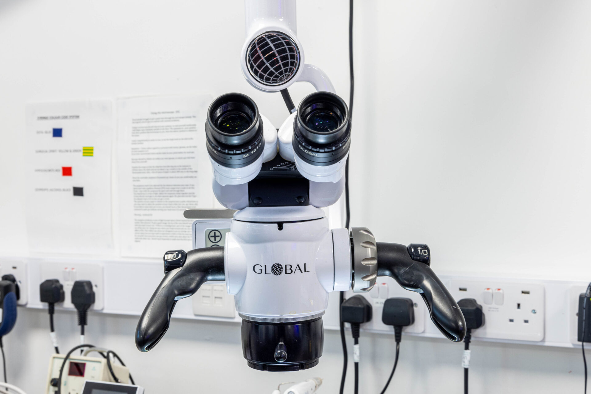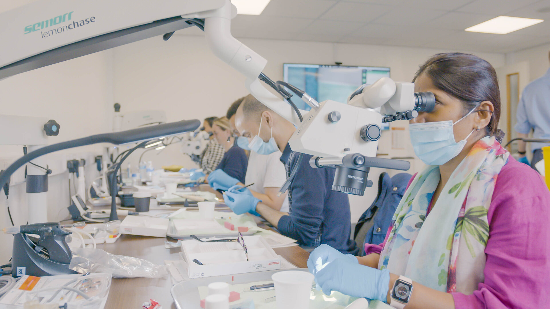
We are absolutely delighted to announce the YEARLY winner of our simplyendo competition ‘Capturing Endodontic Excellence’ A huge congratulations to Justin Underwood on his case.
All the simpyendo tutors agreed this was a very well executed case.
Clinical Case with Persisting Disease
Periradicular Microsurgery (Lower Molar)

Patient Details: Female, 43
3 Appointments: Consultation, Apicectomy, Review
MClinDent Programme: Year 2
Location: Guy’s Hospital, Endodontic Department, Floor 22
Persons Present: Clinician, Qualified Nurse, Patient (Alone), Clinical Supervisor (Consultant & Specialist)
Reason for Referral: A Specialist Endodontist in London Referred the Patient to Guy’s Hospital after their LR6 Retreatment had a Persistent Sinus Tract.
Presenting Complaint
- The Patient is Not in any Pain nor Discomfort since the Retreatment
- An Intermittent Swelling Develops on the Gum which Drains Pus
- The Patient can Eat as Normal
History of Presenting Complaint
- The Primary Root Canal Treatment was Completed by a GDP in Lithuania approximately 15 Years Ago.
- During this time, the Tooth had been Asymptomatic.
- Her GDP Referred the LR6 for Retreatment to a Local Specialist Endodontist who Treated it in September 2021.
- Retreatment was Performed in 1 Visit (under Rubber Dam and Operating Microscope) & Reviewed 6 months later.
- After Retreatment the Patient reported Several Episodes of Well-Localised Moderate Intensity Dull Pain to the LR6 and her GDP Prescribed 500mg Amoxicillin TDS 7 days, which was Effective.
- The Onset of Pain didn’t follow any Pattern.
- The Patient reports that a Draining Infection was Present Before Retreatment and Never Resolved.
- The patient couldn’t remember when they last took Painkillers
Relevant Medical history
- Medical Conditions: Gilbert Syndrome, Bartholin Gland Surgery.
- No Medications.
- Patient confirmed no Allergies including Household Bleach, Latex, Nickel, Titanium.
Dental History
- Diet History: Low Sugar Intake.
- Brushes x2 with the Bass Technique & Electric Toothbrush, Rarely uses Mouthwash, Regular Tepe Use
- Regular Dental Attendee
- LR6 Holds High Strategic Importance to the Patient
- Low Dental Anxiety
- No Previous Dental Trauma Reported
- History of Orthodontic Treatment with Invisalign by a Private GDP
- No Known Bruxism or Clenching
Social history
- Non-Smoker
- Drinks 3 Units of Alcohol / Week
- Strategic Procurement Business Partner
- Lives Locally in London. No Preference for Attending Appointments
Extra-Oral Examination
- No Facial Asymmetry
- No TMJ Clicking
- No Palpable Lymph Nodes
- No Abnormal Neck Swelling
- Healthy Muscles of Mastication
- Competent Lips
Intra-Oral Examination:


- General State: Fully Dentate, Mildly Restored, Well-Maintained Dentition
- Gag Reflex: Not Hypersensitive
- Mouth opening: 3 Fingers Width
- No Visible Caries

Periodontal
Excellent Oral Hygiene
No Supra or Subgingival Calculus
Healthy Periodontium
Generalised Soft tissues: Healthy
Localised Soft Tissues around LR6 No Swelling/ No Sinus Tract / No Erythema:
Dental Chart

Dental Examination
- Tooth Wear: Basic Erosive Wear Examination (BEWE): 000/000
- LR6 shows No Discolouration
- LR6 shows No Visible Fractures
- LR6 shows No Gingival Recession
- Sound and Extensive MOD Composite Restoration. Thin Buccal and Lingual Walls
- From an External inspection, there appears to be Sufficient Tooth Structure present to consider LR6 as Predictably Restorable for an Onlay Restoration
Occlusion
- Static: Class 1 Evenly Distributed Malocclusion
- Dynamic: Left and Right Canine Guidance
- Opposing: UR6 is present
- Adjacent: LR5 and LR7 are both present
No Evidence of Parafunctional / Occlusal Trauma.
Special Tests
- EPT LR7 & LR5: Positive Responses (Scores 23 and 30 Respectively).
- Cold Sensibility Test (Endo Frost): LR7 and LR5. Both Normal Positive Response within 3 Seconds (Not Hypersensitive, Non-Lingering)
- LR6 has No Buccal Palpation Tenderness, nor Lingual Palpation Pain
- Normal and Healthy Periodontal Probing Depth with No Deep Isolated Periodontal Pockets
- No Mobility
- No Tenderness to Percussion
- No Pain to Biting on Cotton Roll Biting
Risk Factors
- Oral Cancer Risk: Low
- Periodontal Risk: Low
- Caries Risk: Low
- TSL Risk: Low
Radiographic Examination
All radiographs followed ALARP principles (As Low As Reasonably Practicable)
Justification: Assessment of:
- Periapical Pathology for Endodontic Diagnosis,
- Root Canal Anatomy
- Restorability and Case Difficulty to aid Treatment Planning
- Prognosis

Grade A:
- Radiographic Report.
- Teeth Present: LR8, LR7, LR6, LR5, LR4
- Partially Imaged: LR7, LR4
- 5% Horizontal Bone Loss
LR7:
- Well Adapted M.O Composite Restoration without Caries
- Healthy Apical PDL
LR6:
- Extensive Coronal Restoration with Acceptable Marginal Integrity
- No Caries Present.
- Open Contact Point Mesially
- Optimal GP Tapers in Both Roots and within 2mm of the Radiographic Apices
- GP has Good Homogeneity without Evidence of Voids
- No Evidence of Iatrogenic Mishaps
- Extensive and Well-Defined Periapical Radiolucency >5mm along the Distal Root extending to the Mid Root Level, <5mm in the Mesial Root.
- No Evidence of Resorption
LR5:
- Unrestored with Possible Enamel Caries Distally.
- Healthy Apical PDL
Cone Beam Computer Tomography (CBCT)
Justification: ESE guidelines: Pre-surgical Assessment prior to Complex Periradicular Surgery
Report: Grade A


Separate Well-Defined, Non-Corticated, Unilocular, Uniformly Radiolucent Lesions Extending from the Distal Aspect of the Mesial and Distal Roots
-

- Coronal View Showing Two Mesial Root Fillings with 2 Separate Foramena
-

- Coronal View Showing Two Distal Root Fillings with 1 Common Foramen The DB Root is Ledged Halfway
Definitive Diagnosis:
- Periodontal Health
- LR6: Chronic Apical Periodontitis Associated with a Previous Rooted Treatment
Treatment Options Offered
- LR6: Another Non-Surgical Retreatment
- LR6: Periradicular Microsurgery
- LR6: Extraction and Accept Gap.
- Extraction & Replace Maryland Bridge (Abutments LR7 & LR5) & Replace w. a Definitive Maryland 6/12
- Extraction & Replace with Removable Prosthesis: Partial Acrylic Denture + Reline + Replacement in 6 months.
- Extraction & Replace with Removable Prosthesis: Partial Chrome Denture + Reline + Replacement in 6 month
- Extraction & Revisit in 3/12 for a Future Implant Subject to Investigations by an Experienced Implantologist
- Decline Treatment and Monitor Radiographically
Dentist’s Recommendation:
- LR6: Periradicular Microsurgery
- Cuspal Coverage Restoration – Onlay
Patient’s Decision: Patient Agreed.
Consent
- Advantages, Disadvantages, Risks, Warnings and Prognosis regarding Periradicular Microsurgery were discussed (Appendix 1)
- Advantages, Disadvantages, Risks, Warnings & Prognosis regarding Extraction were discussed (Appendix 1)
- Advantages, Disadvantages and Warnings & Prognosis regarding No Treatment were discussed (Appendix 1)
- Patient demonstrated Capacity in understanding everything discussed.
- Informed Valid Consent Achieved Verbally and Documented in Patient Records.
Perceived Difficulty of the Endodontic treatment: Class III (Highly Complicated)
Prognosis:
Endodontic Fair
Prosthodontic: Favourable
Periodontal: Favourable
Treatment Plan – Endodontic Department
- LR6: Periradicular Microsurgery
- 1 Week Review: Suture Removal
- 12 Month Review
Treatment Plan – General Dentist Practitioner
- LR6: Indirect Extra-Coronal Restoration – Onlay.
- LR5: Monitor Enamel Caries
- Diet Advice
- Fluoride Advice (+/- Fluoride Varnish and/or Prescription Fluoride Toothpaste as per BNF)
- Routine Radiographs (as per FGDP Guidelines based on Caries Risk Assessment)
Description of Treatment
LR6 Apicectomy was carried out under an Operating Microscope according to pre-defined protocols as per Appendix 12,
Specific Treatment Details
- The Treatment was Performed with the Patient Biting on a Saline Gauze with their Head Turned Sideways
- 3-Sided Papilla-Sparring Periosteal Flap was raised
- The Relieving Incision was Distal to the LR7 extending Mesially to the LR5 (which only extended half way to protect the Mental Nerve)
- No Cracks/Root Fractures Identified during the Apicectomy
- Difficult Access, especially in the Lingual Canals
- Difficult Suturing
- The Flap was Sutured with Ethilon 5/0 Non-Absorbable Monofilament Sutures
- At the 7-day Review, the Patient Reported having Extensive Post-Operative Swelling and Bruising lasting 5-6 days & Pain Lasting 3 days requiring 4-6 Hourly 400mg Ibuprofen & 1000mg Paracetamol
- Clinically, Tissue Fluid was Oozing from the site but No Pain, Bad Taste, Nor Bad Odour present.
-

- Mid Treatment Radiograph. Grade A. Sufficient Root Resection has occurred.
-

- Mid Treatment Radiograph. Grade A. Good Retro-fill in All 4 Canals
Post-Operative Advice
Sterile Gauzes, Cold Pack and Written Post-Operative Aftercare Advice was given to patient as per Appendix 13
12 Review Appointment

Healing on both Mesial and Distal Roots.
- The Patient reported No History of Symptoms since the LR6 Apicectomy.
- Clinically, the Soft Tissue were Healthy with No Evidence of Erythema, Sinus tract nor Swelling
- LR6: No TTP, Normal Mobility, No Pain on Buccal/Lingual Palpation and No Deep Isolated Pocketing.
- Periapical Radiograph Examination revealed Excellent Bony.
Written Communication
3 letters were posted to the GDP and Patient involving a:
- Consultation Summary,
- Treatment Summary
- 12 Month Review Summary.
Discussion
The most ideal management of iatrogenic errors would be its prevention, however, a large scope of an endodontist’s practice is rectifying other dentist’s errors duting retreatments and performing periradicular microsurgery when necessary. Thankfully, the success rate of surgical endodontic treatment with modern techniques is 89% (Tsesis et al 2015).
Mandibular molars present a technical challenge when performing periradicular microsurgery due to its proximity to the ID nerve and difficulty in accessibility. The relationship between the apices of both the first and second molars to the inferior alveolar nerve is generally between 3.5–5.4 mm (Littner. M, 1986). In this case, the pre-operative CBCT confirmed that the distance from the inferior alveolar canal was 6 mm, which is a safe margin for this surgery.
Diligence was needed in the relieving incision to avoid the mental nerve. A full‑thickness mucoperiosteal flap could have been selected, however a papilla-based flap was chosen to minimise possible gingival recession which could result in dentine hypersensitivity of the adjacent teeth.
On flap reflection there a was a natural bony breach in the buccal bone which assisted with the orientation. The osteotomy was enlarged to visualise both roots. During the treatment, the dental assistant found her role in this surgery quite challenging due to difficulties flap retraction due to the surgical access.
The rear-vented fast handpiece with a 555 burr was at full reach during the root resection making visualisation tricky. Due to the existing ledge in the DB canal, the root resection had to be made higher in the root than ideal. After the root resection and retro prep, MTA was used due to its physical and biological properties. This includes an excellent seal, good marginal adaptation, reduced microleakage, high biocompatibility, bioactive & antimicrobial properties (Wahengbam et al, 2014). As the flap design was entirely buccal, suturing was technically challenging and time consuming.
During this treatment the patient had to maintain their head in a fixed position to the side to assist with my visualisation. Overall I am delighted with the quality of this surgery, I feel that the tooth has a reasonable prognosis, and the patient now needs to see their general dentist for an extra-coronal restoration to protect the cusps.
References
Tsesis, I., Rosen, E., Taschieri, S., Telishevsky, Strauss, Y, Ceresoli, V., Del Fabbro, M. (2013) Outcomes of surgical endodontic treatment performed by a modern technique: An updated meta‑analysis of the literature. Journal of Endodontics, 39, 332‑9.
Littner, M., Kaffe, I., Tamse, A., Dicapua, P. (1986) Relationship between the apices of the lower molars and mandibular canal – A radiographic study. Oral Surg Oral Med Oral Pathol, 62, 595‑602.
Wahengbam, B., Wahengbam, P., Tikku, A (2014) A simplified technique of orthograde MTA obturation on the elected canals of posterior teeth: Two case reports. Journal of Conversative Dentistry, 17, 80‑4.